

What is all the 'FUS' about with focused ultrasound for Parkinson's disease?
By Michael S. Okun

A recent review in JAMA Neurology shows uses of FUS by Vibhor Krishna in 2018.
A day rarely goes by without a paper, advertisement or video ‘popping up’ about focused ultrasound therapy (FUS) for Parkinson’s disease or essential tremor. Just this past week, another article appeared in the New England Journal of Medicine on its potential use in Parkinson’s disease. So, what is all the ‘FUS’ about? In this month’s blog, I will ‘dive’ into the topic and explore FUS basics as well as the advanced uses of the FUS technology. I directly address the question on everyone’s mind? What are the differences in FUS compared to conventional lesion based therapies and to DBS. Finally, I will address the importance of a ‘shared decision making approach’ for FUS therapy. Since there is a lack direct comparative trials of FUS to DBS, I argue that in practice, you should employ a shared decision making process when deciding on the ‘right procedure for the right person.’
What is deep brain stimulation and how is it different from focused ultrasound?

A review of deep brain stimulation appeared in the NEJM in 2012.
DBS is a medical procedure where doctors insert wires into the brain and connect them to a ‘pacemaker’ like device. The ‘pacemaker’ sends signals to the brain circuits in an effort to change the ‘conversation’ occurring within your brain.’ DBS addresses malfunctioning circuits in diseases such as Parkinson’s or essential tremor; DBS disrupts these circuits. DBS involves drilling a ‘dime sized’ hole in the skull. Sometimes recordings are performed by clinicians and physiologists to confirm the final position of the DBS lead.
Focused ultrasound (FUS) in contrast, is a procedure performed by a clinician from ‘outside’ the brain. The FUS technology uses sound waves to make the brain lesion while you lay within a MRI scanner. A practice test lesion’ can be placed to assess for potential side effects. These test lesions have been helpful, however they do not always predict long-term side effects.
The final FUS lesion (hole) is made in many of the same structures which are stimulated when using deep brain stimulation (thalamus, STN, GPi).

Journal of Neurosurgery review by Quadri 2018 shows a nice cartoon on how FUS works.
What are the main differences between focused ultrasound and deep brain stimulation you should consider when making a decision?
Here is a list of some of the key points I talk about with patients and families who are interested in deep brain stimulation vs. focused ultrasound.
Deep Brain Stimulation:
A small hole is drilled in the skull.
Recordings can be performed to map the precise area where a DBS lead can be implanted.
Bilateral procedures can be performed safely.
The leads can be removed, however because the leads are passed through the brain, they will create minor damage to the brain tissue.
‘Millimeters’ matter in the placement of the DBS lead and the outcome of the procedure.
Mapping the brain ‘from the inside’ provides more precision than FUS or radiosurgery.
The DBS devices can be ‘tuned’ and programmed (over time) to maximize benefits and to minimize side effects.
There are more clinical visits required for DBS programming and management, when compared to a lesion therapy.
There are more long term side effects reported from using DBS including potential infections of the device.
There are more brain bleeds associated with DBS therapy.
Focused Ultrasound
No drilling or burr hole.
The procedure is performed from ‘outside the brain’ and therefore it is less precise than deep brain stimulation.
The procedure is less precise than brain mapping which is performed prior to open brain lesioning procedures.
There is a higher potential for a permanent side effect with ultrasound therapy compared to DBS, especially if the intended target is missed.
Less follow-up is required with focused ultrasound. No device adjustments are required for FUS.
At the current time, focused ultrasound is usually only performed on ‘one side of the brain (unilateral) because of the well known adverse effects from placing bilateral brain lesions.
Less infections and also there are no long term device related issues with focused ultrasound treatments.
Some folks cannot have focused ultrasound because of skull thickness.
Frail, elderly patients with many co-morbidities and/or on blood thinners who are at high risk for open brain surgery could be ideal candidates for focused ultrasound therapy.
What other ways can you make a ‘hole in the brain’ besides focused ultrasound therapy?
It turns out that we have been making ‘destructive lesions’ in human brains for 75 years in procedures referred to thalamotomy, subthalamotomy and pallidotomy. We name the procedures based on the structures which are ablated. The two most common ways
High intensity ultrasound was introduced in the 1940’s and 1950’s as a treatment for a variety of brain disorders. The recent ‘rebirth’ of FUS has generated a lot of enthusiasm and excitement. The ‘repackaging’ of this approach has included combining ultrasound with high field MRI scanning.

There is one other technique called radiosurgery (usually by a gamma knife) which can also be used to make a lesion in the brain. In radiosurgery, the surgeon aims X-ray beams at the brain and destroys tissue. Both radiosurgery and ultrasound destroy the tissue, howeverwhen using ultrasound there is an option to apply a ‘test-lesion’ prior to placing a permanent one. Radiation therapy has another disadvantage when compared to ultrasound. Using X-rays can lead to necrosis of brain tissue (dying), and uncontrolled growth of radiation induced brain lesions. These lesions can in some cases expand in size, lead to delayed complications, and in one case when too much radiation was applied, it resulted in a death (Parkinson’s Foundation blog by Dr. Okun).
What is known about focused ultrasound for essential tremor?

Jeff Elias and colleagues published a nice series of patients who had focused ultrasound thalamotomy for essential tremor.
The first FDA approved indication for focused ultrasound was for medication resistant essential tremor. The procedure is performed on one side of the brain because of the risks of speech and cognitive side effects. Jeff Elias at the University of Virginia and colleagues have recently published several papers on the safety and effectiveness of a thalamic lesion which is placed to treat essential tremor using focused ultrasound technology. Here is one recent paper from the New England Journal of Medicine on FUS for ET. This group also published a paper on focused ultrasound to address tremor in Parkinson’s disease. The most common side effects from ultrasound guided thalamotomy are typically slurred speech and unsteady gait.

Outcomes from the Elias Essential Tremor NEJM study.
Thalamic lesions can be an excellent approach for treatment of upper extremity tremor in Parkinson’s, however thalamic lesions are not as effective as other targets (STN and GPi) on symptoms like rigidity, bradykinesia and dyskinesia. Here is a recent article by Bond and colleagues in JAMA Neurology showing benefits on tremor in Parkinson’s disease.
We have much less long-term safety and efficacy data on FUS when we compare to DBS or to conventional lesioning procedures.

Bond and colleagues in 2017 showed focused ultrasound could be good for Parkinson tremor. Usually not employed for other features of Parkinson.
What is known about focused ultrasound subthalamotomy?

One research group who has been prolific in subthalamic lesioning by ultrasound technology has been the Spanish scientists Obeso and Martinez-Fernandez. Most of the available data on FUS STN lesions has been collected on unilateral procedures and in folks with highly asymmetric Parkinson’s disease. The early available data has revealed benefits and most concerns have mainly been centered on the tiny size of the STN brain target (150 cubic millimeters or the size of a squished pea) and ‘unintended consequences’ of missing the region and accidentally targeting another brain area. The other concern has been ‘hemiballismus’ which is a side effects that is common in STN region strokes but can also be observed following subthalamotomy. Hemiballismus is most commonly a ‘flailing movement of the arm and/or leg.’

Martinez-Fernandez pioneering work on subthalamic lesions in Parkinson’s using a focused ultrasound methology.
What do we know about focused ultrasound pallidotomy?

Much of the recent media attention on focused ultrasound has been driven by the recent pallidotomy publication which appeared in the New England Journal of Medicine. In this study, folks with Parkinson’s disease and dyskinesias, or alternatively those with motor fluctuations and motor impairment, when in the off-dopaminergic medication state, were randomized to focused ultrasound versus a sham procedure. The results revealed benefit in 69% in FUS group; and 32% in the control group. There were 30/39 patients who maintained a 12 month response. Adverse events in the active group were mainly dysarthria, gait disturbance, loss of taste, visual disturbance, and facial weakness. TTwenty three percent of patients in the FUS group did not maintain a response at 12 months. Visual adverse effects will be important to track because of the lesion’s proximity to the optic track and due to the decreased precision when using a tool like FUS (see NEJM Journal Watch for full summary).

The importance of shared decision making when choosing FUS vs. DBS vs. a conventional lesion based therapy
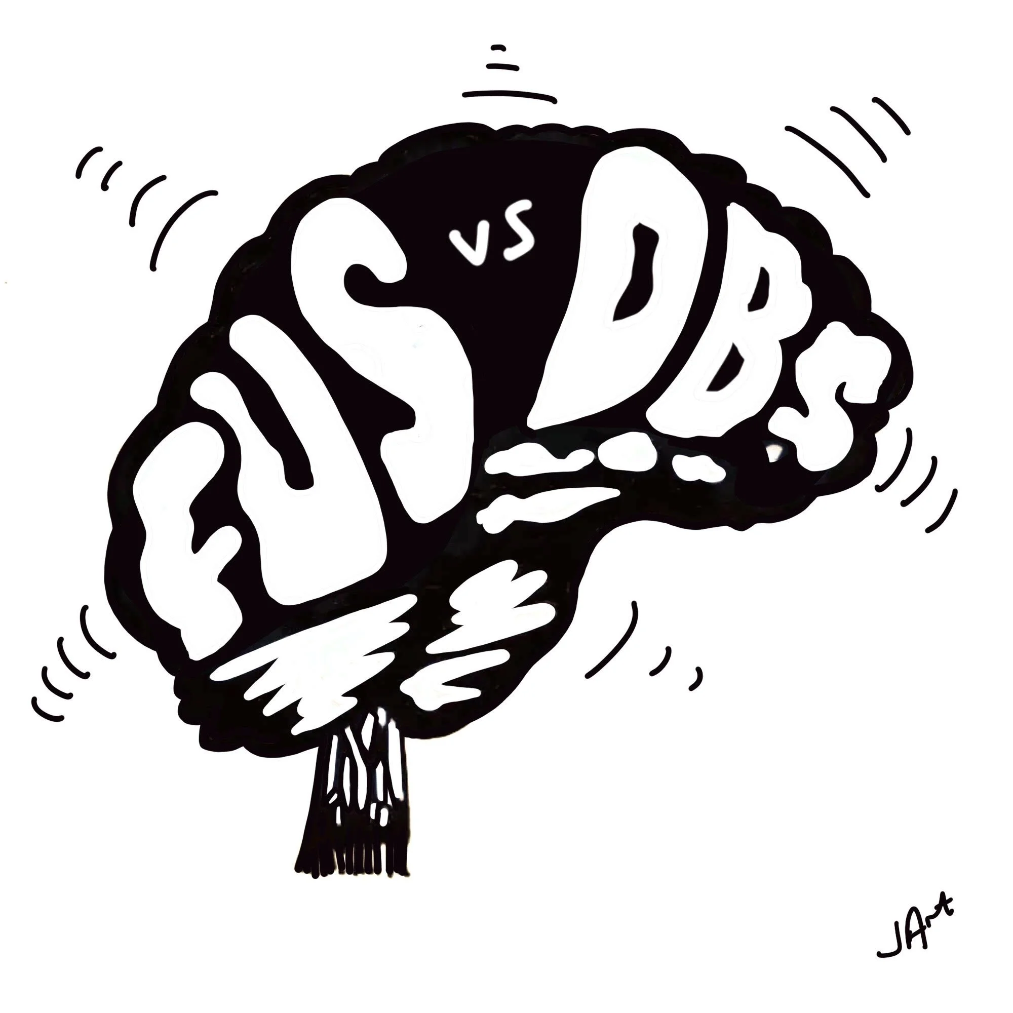
Jonny Acheson drew this great pic to encapsulate the DBS vs. FUS conundrum within each person’s mind.
Critical to deciding on a surgical therapy for an individual patient, is shared decision making. The risks and benefits of each approach should be discussed and adequate time should be budgeted for the discussion; as the topic is complex. The clinician and the patient should cover the following key questions.
Do i need one sided (unilateral or bilateral) surgery?
What symptom or symptoms do you seek to improve, and which therapy will best address your need.
How do you feel about a brain lesion versus an electrical device?
Are you excited about the ability to ‘tune your device’ to symptoms over time or do you prefer a ‘one and done approach.’
If you prefer a lesion based therapy, would you favor an open surgical technique if the outcome(s) were overall better?
If you are considering the thalamic target for Parkinson’s disease make sure you discuss the possibility that this target may not address the other features of the syndrome (stiffness, slowness, dyskinesia).
Are you interested in the future possibility of a neuromodulation technique which may possibly be programmed to your specific brain signals and symptoms.
Are you on blood thinners or do you have any medical reasons which would place you in a high risk category for an ‘open surgical technique.’

Parkinsonsecrets.com blog editor Michael S. Okun. Dr. Okun works with Drs Subramony and Acheson on the content for this site.

Dr. Jonny Acheson is an artist, doctor and person with Parkinson and the artist for this blog.